Home » Uncategories » Pictures Of Muscles And Bones : AWESOME ORIGIN/INSERTION VISUAL (anatomy-muscular system ... - The talus bone is the second largest bone in the entire foot, and unlike the rest bones, there is no attachment of muscles.
Pictures Of Muscles And Bones : AWESOME ORIGIN/INSERTION VISUAL (anatomy-muscular system ... - The talus bone is the second largest bone in the entire foot, and unlike the rest bones, there is no attachment of muscles.
Pictures Of Muscles And Bones : AWESOME ORIGIN/INSERTION VISUAL (anatomy-muscular system ... - The talus bone is the second largest bone in the entire foot, and unlike the rest bones, there is no attachment of muscles.. This anatomical atlas was especially designed for a specific public (radiologists. 31 full pdfs related to this paper. Human arms anatomy diagram, showing bones and muscles while flex human arms anatomy diagram, showing bones and muscles while flexing. Skull sutures, temporomandibular, shoulder, elbow, wrist, hip, knee, and ankle joints It forms the lower part of the ankle (formed collectively by the tibia, fibular, and talus bones).
Bones and muscles of the foot. Muscles, joints, and bones work together so your body can move harmoniously. The smaller bone that runs alongside the tibia (fibula) and the kneecap (patella) are the other bones that make the knee joint. The shoulder is the region where the upper limb is attached to the trunk. Collection film x ray shoulder radiograph show shoulder dislocation and bone broken (neck of humerus fracture) from accident highlight on arrow point.
Treating Levator Scapula Muscle (Shoulder Muscle) Pain from fthmb.tqn.com It forms the lower part of the ankle (formed collectively by the tibia, fibular, and talus bones). Many of the muscles that move the fingers and thumb originate in the forearm. A short summary of this paper. Primarily, it functions to transmit your body's weight to the foot through the talocalcaneonavicular joint. The basics on muscles, bones, and joints. The shoulder is the region where the upper limb is attached to the trunk. Bones and muscles of the foot. On the other hand, the insertion is where a tendon attaches that muscle to the *more* movable bone.
The forearm's ulna and radius support the many muscles that manipulate the bones of the hand and wrist.
The musculoskeletal system consists of the body's bones, muscles, tendons, ligaments, joints, & cartilage. The basics on muscles, bones, and joints. The bones of the hand and wrist provide the body with support and flexibility to manipulate objects in many different ways. See more ideas about muscle anatomy, leg muscles anatomy, anatomy. The fleshy, thick part of the muscle is called its belly. This anatomical atlas was especially designed for a specific public (radiologists. Key facts about the main bones, joints and muscles of the body; Primarily, it functions to transmit your body's weight to the foot through the talocalcaneonavicular joint. 2 d digital illustration, on white background. Then, when the movement is completed, the flexor relaxes and the extensor contracts to extend or straighten the limb at the same joint. When there is damage to one of the structures that surround the knee joint, this can lead to discomfort and disability. The image below shows the bones of the hand from the back side. Muscles can pull bones, but they can't push them back to the original position.
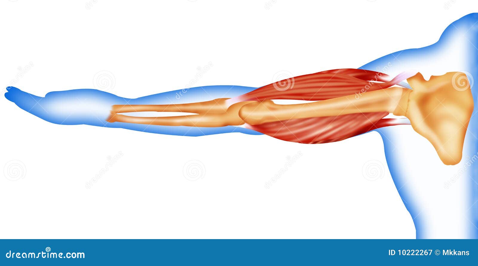
Tendons connect the knee bones to the leg muscles that move the knee. Bones of the upper and lower limbs and the shoulder and pelvic girdles main joints: We'll go over the bones, joints, muscles, nerves, and blood vessels that make up the human arm. The shoulder is the region where the upper limb is attached to the trunk. Both, arm bones and muscles work in coordination with each other to makes various functions of the hand possible.
Body muscles and bone stock illustration. Illustration of ... from thumbs.dreamstime.com September 23, 2019 edited by dr. The forearm's ulna and radius support the many muscles that manipulate the bones of the hand and wrist. The shoulder is the region where the upper limb is attached to the trunk. Tendons connect the knee bones to the leg muscles that move the knee. The musculoskeletal system consists of the body's bones, muscles, tendons, ligaments, joints, & cartilage. The knee joint is a complex structure that involves bones, tendons, ligaments, muscles, and other structures for normal function. The image below shows the bones of the hand from the back side. Secondarily, it protects the spinal cord (which is the extension of the brain) and all of the nerves that branch from the spinal cord.
Find diagnosis, treatment, and prevention information on more than 20 different muscle and bone diseases and conditions affecting the musculoskeletal system.
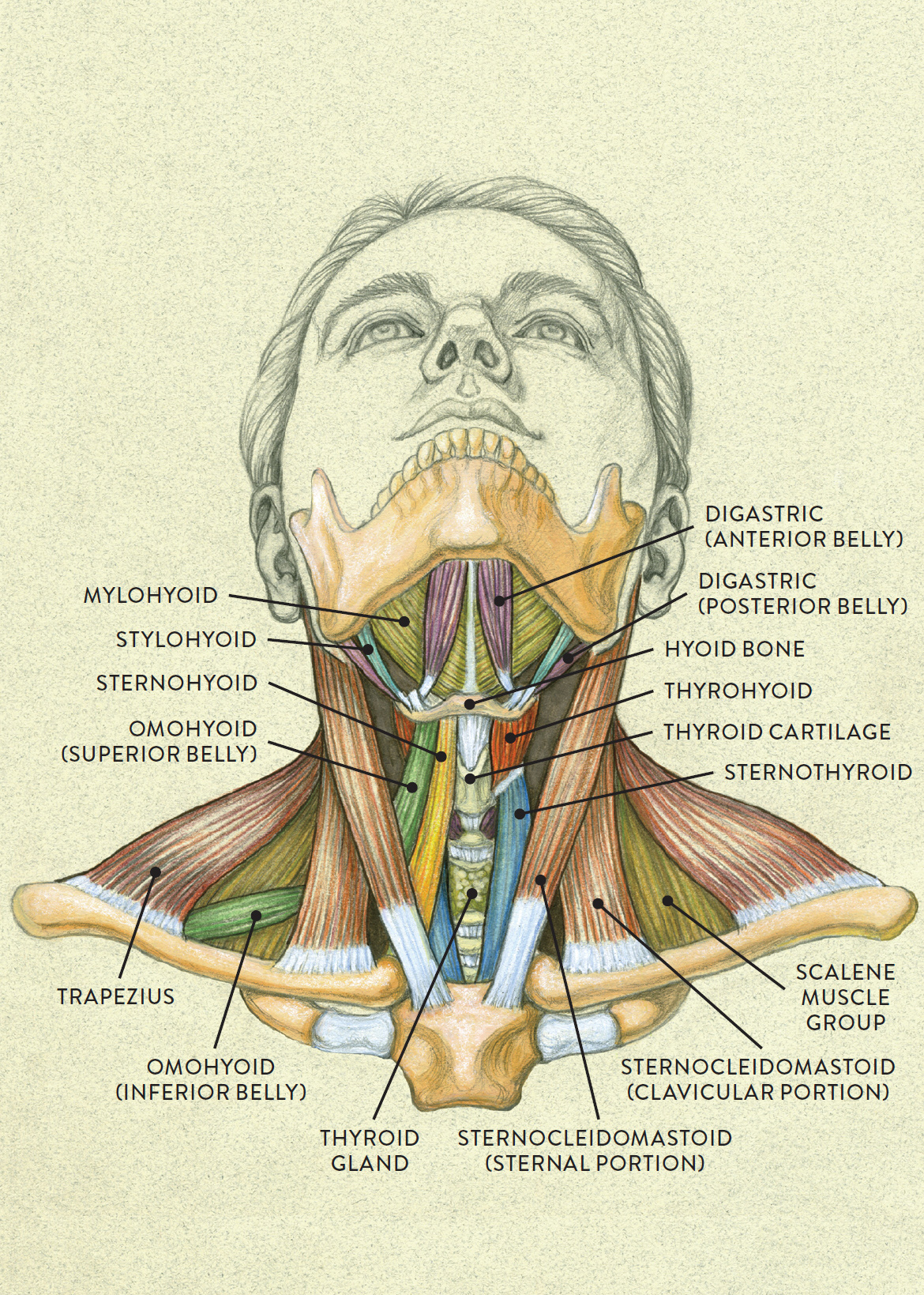
The foot is a part of vertebrate anatomy which serves the purpose of supporting the animal's weight and allowing for locomotion on land. Bone on hand and foot diagram quiz 12 photos of the bone on hand and foot diagram quiz , bone. Then, when the movement is completed, the flexor relaxes and the extensor contracts to extend or straighten the limb at the same joint. Neck anatomy pictures bones, muscles, nerves. A short summary of this paper. Find out how the musculoskeletal system functions — and which medical. Bones of the skull, ribs, vertebral column, sternum, sacrum, coccyx, hyoid bone and auditory ossicles. It is made up of the bones of the skeleton, muscles, cartilage, tendons, ligaments, joints, and. Muscles can pull bones, but they can't push them back to the original position. In humans, the foot is one of the most complex structures in the body. Related posts of neck bones and muscles pictures bone on hand and foot diagram quiz. The humerus is the bone of the arm that articulates with the scapula proximally and with the radius and the ulna distally. 2 d digital illustration, on white background.
Many of the muscles that move the fingers and thumb originate in the forearm. The fleshy, thick part of the muscle is called its belly. So they work in pairs of flexors and extensors. Each hand contains 27 distinct bones that give the hand an incredible range and precision of motion. The purpose of the spine is to support the body so that we can stand upright.
Anterior view of head tilting back from schoolbag.info Primarily, it functions to transmit your body's weight to the foot through the talocalcaneonavicular joint. A short summary of this paper. Test your knowledge of the clavicle, scapula and humerus with our labeled diagram exercises and quizzes! 2 d digital illustration, on white background. Bones of the upper and lower limbs and the shoulder and pelvic girdles main joints: Many of the muscles that move the fingers and thumb originate in the forearm. The arm is one of the body's most complex and frequently used structures. Key facts about the main bones, joints and muscles of the body;
Test your knowledge of the clavicle, scapula and humerus with our labeled diagram exercises and quizzes!
The forearm's ulna and radius support the many muscles that manipulate the bones of the hand and wrist. Many of the muscles that move the fingers and thumb originate in the forearm. The musculoskeletal system consists of the body's bones, muscles, tendons, ligaments, joints, & cartilage. A short summary of this paper. This anatomical atlas was especially designed for a specific public (radiologists. The red lines show where the tendons attach the muscles to the bones. 31 full pdfs related to this paper. The talus bone is the second largest bone in the entire foot, and unlike the rest bones, there is no attachment of muscles. The foot is a part of vertebrate anatomy which serves the purpose of supporting the animal's weight and allowing for locomotion on land. The image below shows the bones of the hand from the back side. Each hand contains 27 distinct bones that give the hand an incredible range and precision of motion. Bones of the skull, ribs, vertebral column, sternum, sacrum, coccyx, hyoid bone and auditory ossicles. Browse 4,015 shoulder bone stock photos and images available, or search for pork shoulder bone to find more great stock photos and pictures.
0 Response to "Pictures Of Muscles And Bones : AWESOME ORIGIN/INSERTION VISUAL (anatomy-muscular system ... - The talus bone is the second largest bone in the entire foot, and unlike the rest bones, there is no attachment of muscles."
/GettyImages-499158129-56a05f075f9b58eba4b0267f.jpg)

0 Response to "Pictures Of Muscles And Bones : AWESOME ORIGIN/INSERTION VISUAL (anatomy-muscular system ... - The talus bone is the second largest bone in the entire foot, and unlike the rest bones, there is no attachment of muscles."
Post a Comment